|
1 Comment
¿Sabías que aproximadamente 1.3 billones de personas viven con algún tipo de discapacidad visual? [1]. Bueno, si no, no me siento tan mal porque esta fue una de las primeras cosas que aprendí cuando visité el póster de Giovanne Diniz y la Dra. Luciane Sita sobre el “Desarrollo de herramientas de aprendizaje de neuroanatomía táctil de bajo costo para estudiantes ciegos y con discapacidad visual”. Siempre trato de mantenerme actualizada sobre las nuevas herramientas que se utilizan para la instrucción inclusiva. Así fue como aprendí sobre el trabajo de Giovanne y la Dra. Sita mientras asistía a los carteles del Tema J en la reunión anual de la Sociedad de Neurociencias (SfN). 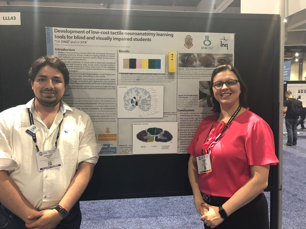 Giovanne Diniz y la Dra. Luciane Sita en su póster durante la reunión anual de SfN. Giovanne y la Dra. Sita son científicos de la Universidad de Sao Paulo, Brasil. El año pasado, cuando comenzaron a impartir el curso de neuroanatomía, se dieron cuenta de que había un alumno ciego en su clase y los materiales de instrucción eran limitados para enseñar a esta población. Luego, comenzaron a identificar herramientas para usar en su instrucción para mejorar la enseñanza para estudiantes ciegos y con discapacidad visual. La mayoría de los cursos de anatomía utilizan ilustraciones, imágenes médicas y cadáveres; aunque existen herramientas didácticas disponibles, son para personas videntes y la mayoría de ellas están en inglés. Así, este equipo decidió desarrollar una herramienta para mejorar la enseñanza de los conceptos de neuroanatomía utilizando materiales de bajo costo, una que pudiera ser accesible e implementada sin dificultad. Entre las herramientas que desarrollaron para la enseñanza de la neuroanatomía se encontraba un espécimen cerebral fijo que tenía las circunvoluciones cubiertas con diferentes telas de textura y marcadas con alfileres de varios tamaños. Este enfoque también se utilizó para enseñar las estructuras internas del cerebro que se presentaron como diapositivas cerebrales dibujadas digitalmente. La implementación de sus herramientas aumentó la participación del estudiante ciego que asistía a su clase. El desempeño de los estudiantes fue similar al de los compañeros videntes y esto los motivó a aumentar el repertorio de estructuras que han desarrollado y a proporcionar esto como un modelo para el uso de otros con estudiantes ciegos o con discapacidad visual. El uso de estrategias táctiles para la enseñanza puede ser difícil, ya que hay varias cosas a considerar, incluidas las necesidades y habilidades de los estudiantes, y las tareas que se implementarán [2]. Las herramientas desarrolladas por Giovanne y la Dra. Sita, que se utilizan para enseñar los conceptos básicos de la neuroanatomía, son una gran estrategia para la inclusión y la instrucción eficaz para los estudiantes ciegos y con discapacidad visual.  Ejemplo de una sección del cerebro dibujada digitalmente y las texturas utilizadas para enseñar sobre estructuras específicas. Referencias * Fui seleccionada como Blogger oficial de la conferencia Anual de la Sociedad de Neurociencias de 2018 y esta fue una de las publicaciones, versión en inglés, de mi blog sobre el tema asignado, el Tema J: Historia y educación de la neurociencia
Did you know that approximately 1.3 billion people live with some form of vision impairment? [1] Well if you didn’t I don’t feel as bad because this was one of the first things I learned when I visited Giovanne Diniz and Dr. Luciane Sita’s poster on “Development of low-cost tactile neuroanatomy learning tools for blind and visually impaired students”. I always try to stay updated on new tools used for inclusive instruction. That is how I learned about Giovanne and Dr. Sita’s work while attending Theme J posters at the annual meeting.  Giovanne Diniz and Dr. Luciane Sita at their poster during SfN’s Annual meeting. Giovanne and Dr. Sita are scientists at the University of Sao Paulo, Brazil. This past year when they started teaching the neuroanatomy course they realized that there was a blind student in their class and the instructional materials were limited to teach this population. Then they began to identify tools to use on their instruction to improve teaching for blind and visually impaired students. Most anatomy courses use illustrations, medical imaging, and cadavers; although there are didactic tools available these are for sighted individuals and most of them are in English. Thus, this team decided to develop a tool to improve the teaching of neuroanatomy concepts using low-cost materials, one that could be accessible and implemented without difficulty. Among the tools they developed for teaching neuroanatomy was a fixed brain specimen which had the gyri covered with different textured fabrics and marked with pins of various sizes. This approach was also used to teach internal structures of the brain which were presented as digitally drawn brain slides. Implementation of their tools increased the engagement of the blind student attending their class. The students’ performance was similar to the sighted peers and this motivated them to increase the repertoire of structures they have developed and to provide this as a blueprint for use of by others with blind or visually impaired students. Using tactile strategies for teaching can be difficult as there are several things to consider including the students’ needs and abilities, and the tasks that will be implemented [2]. The tools developed by Giovanne and Dr. Sita, used to teach neuroanatomy core concepts, are a great strategy for inclusiveness and effective instruction for blind and visually impaired learners. 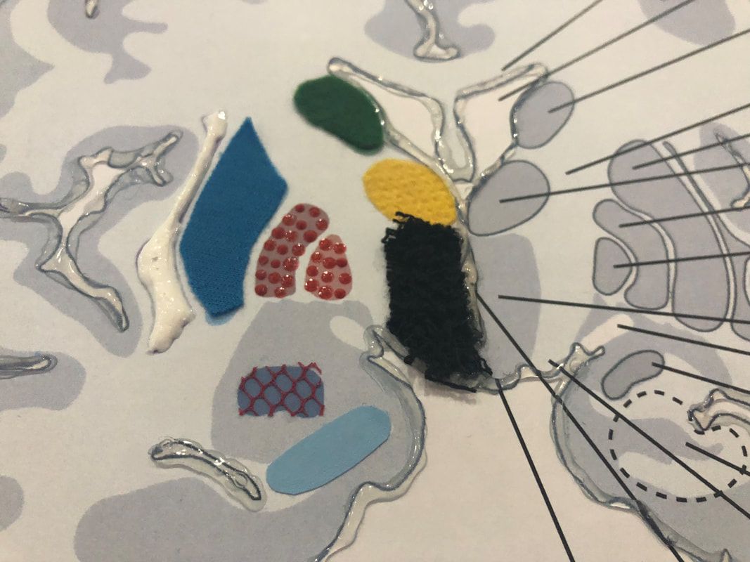 Example of a digitally drawn brain section and the textures used for teaching about specific structures. References *I was selected as an Official 2018 Society for Neuroscience Blogger and this was one of my blog posts about my assigned theme which was Theme J:Neuroscience History and Education.
|